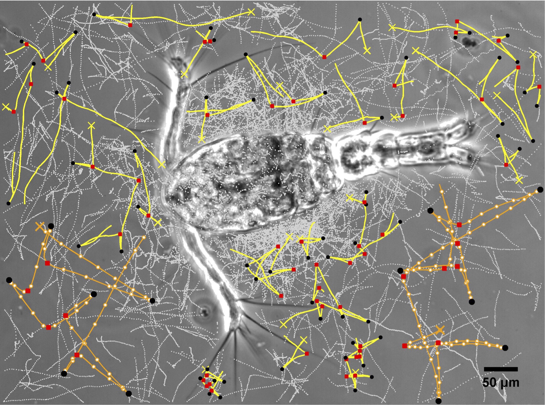
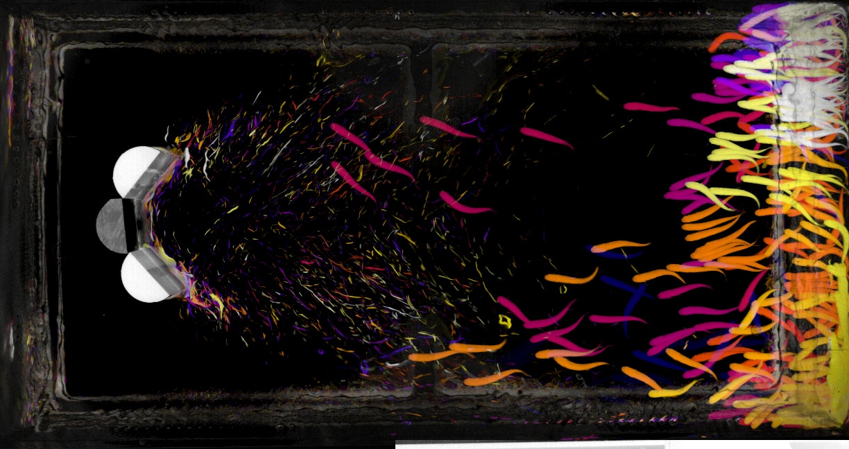
We strive to make micro scale processes and microbial interactions ‘come to life’,
through both experimental and conceptual images and videos.
Feel free to download them and please credit them as indicated.
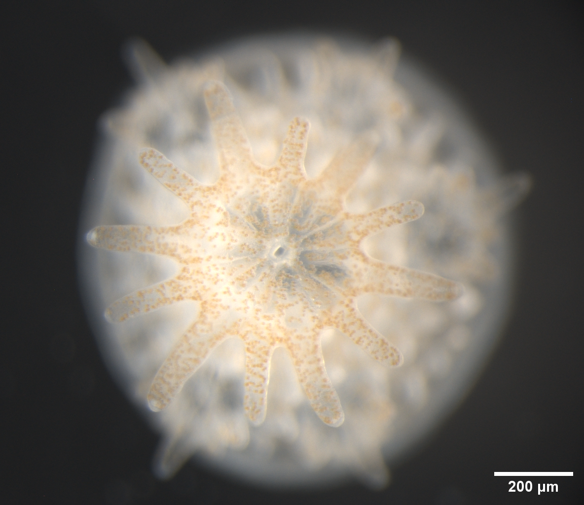
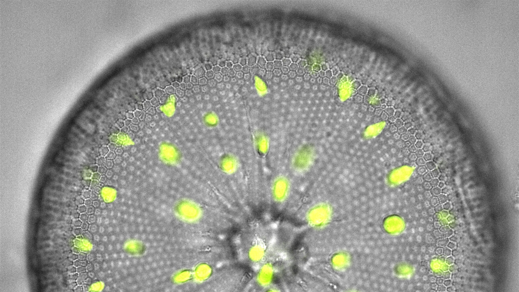
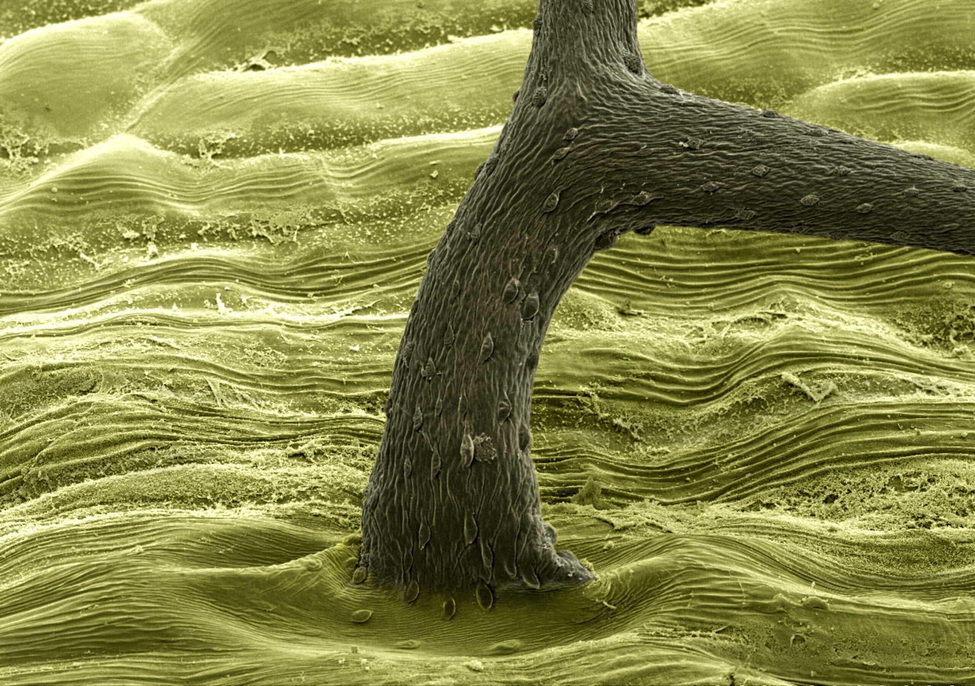
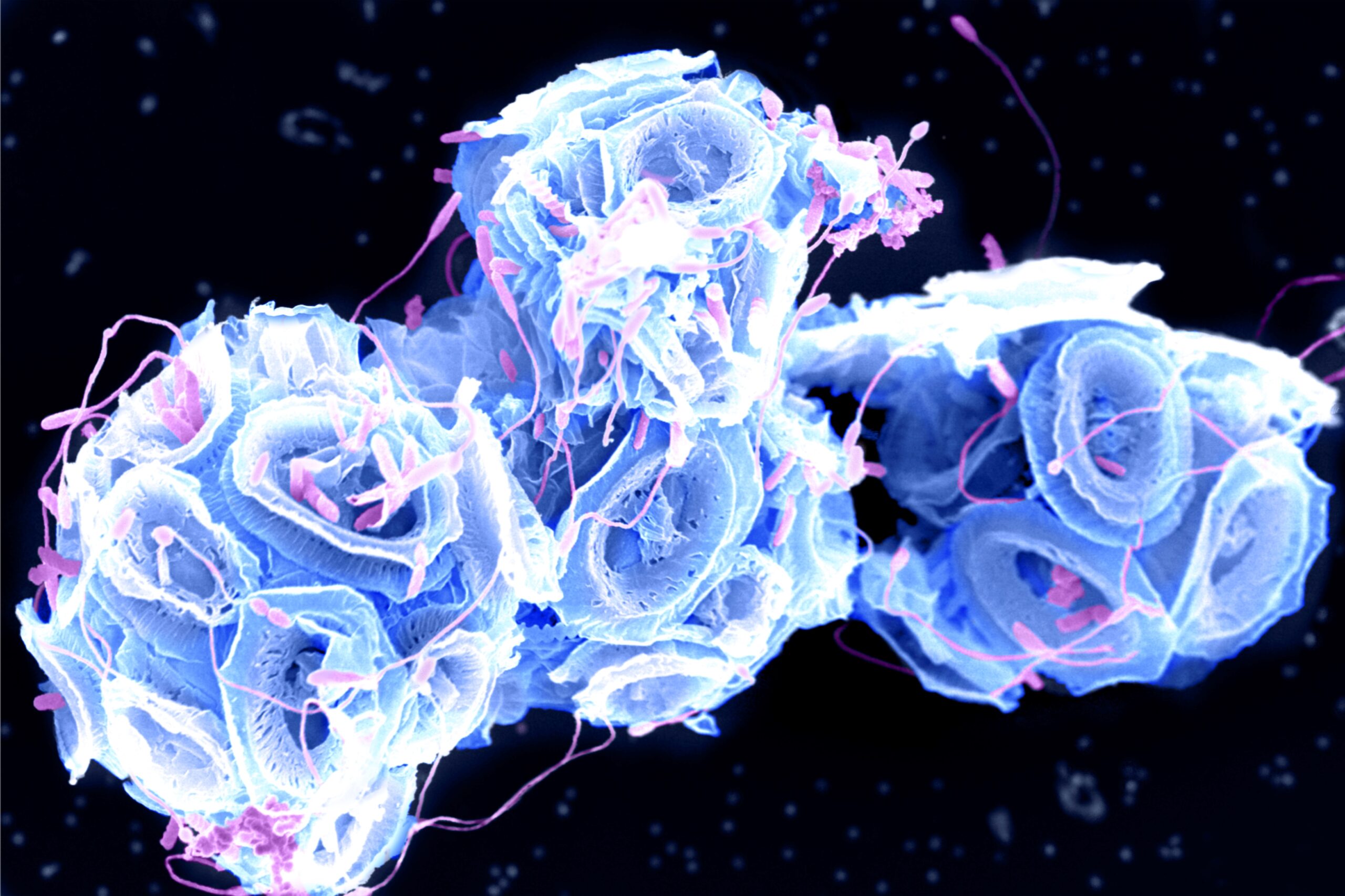
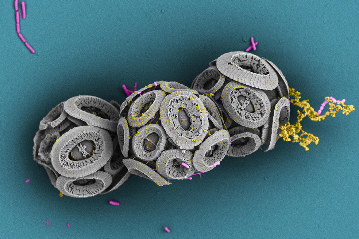
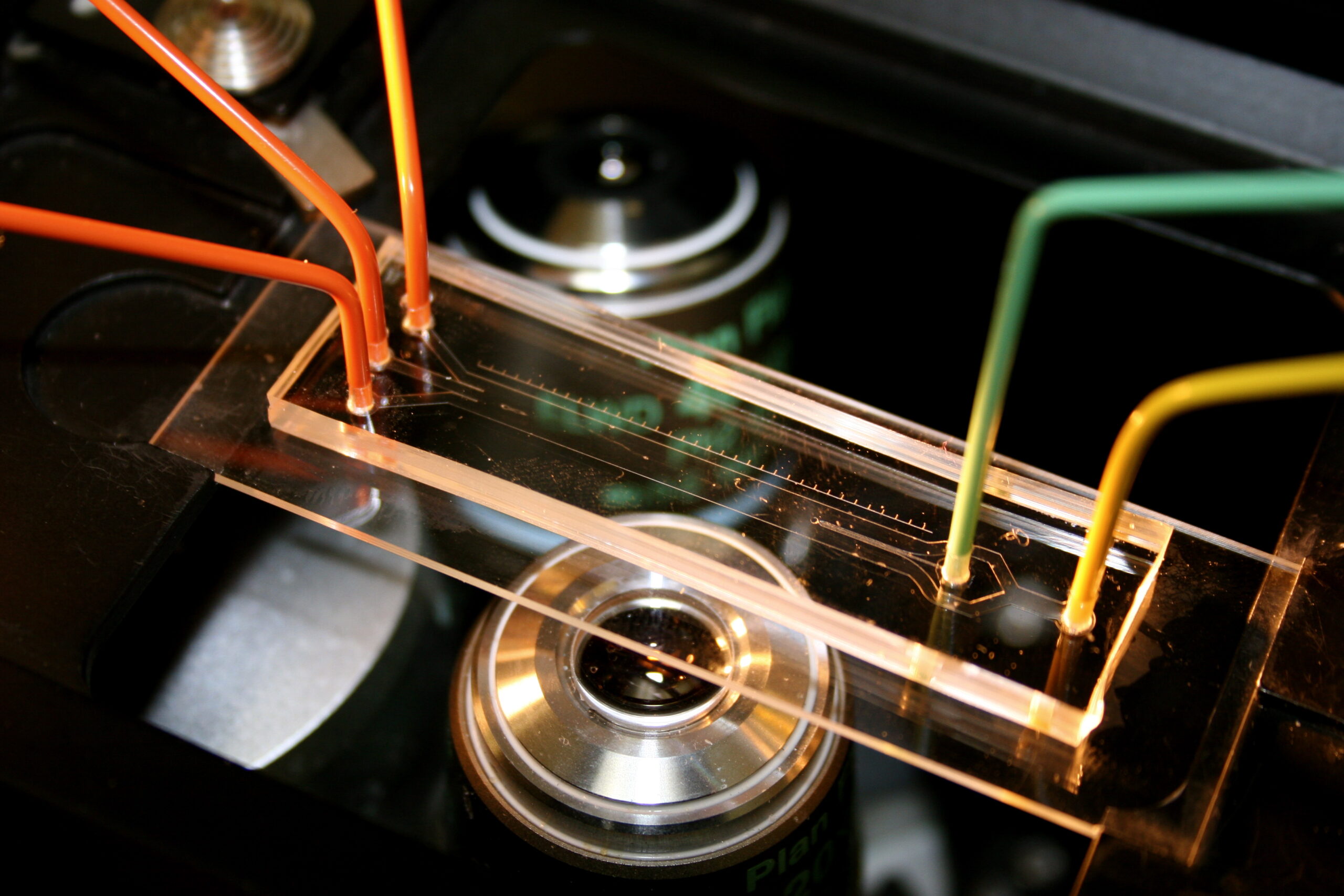
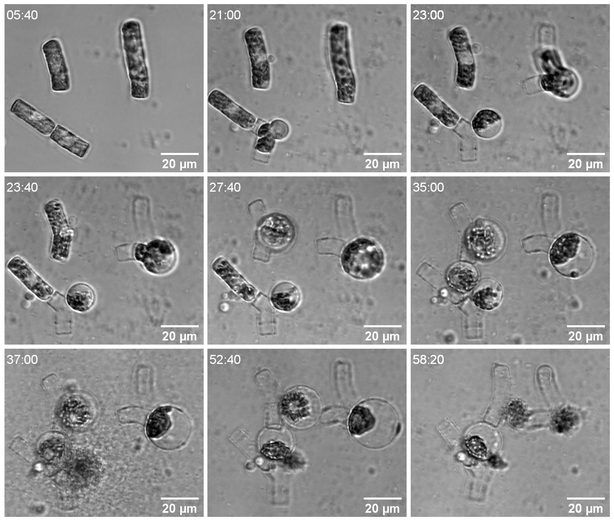
Microscale dynamics of bacteria-spines interactions
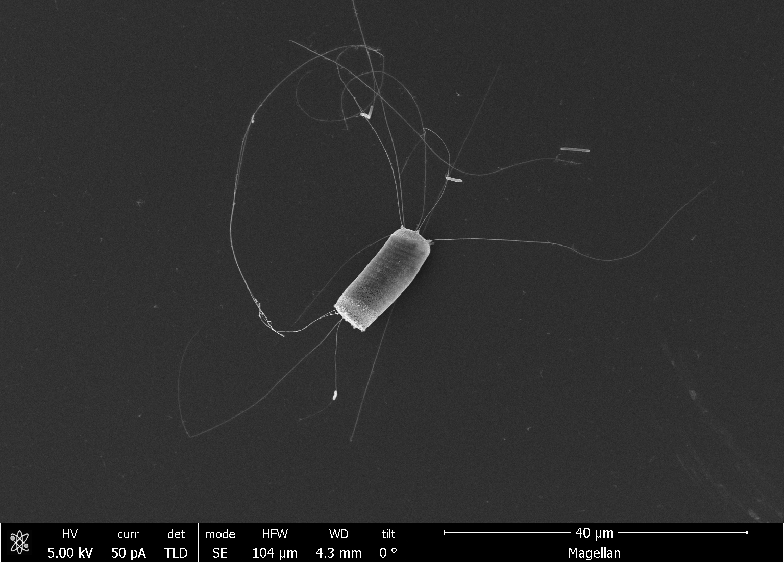
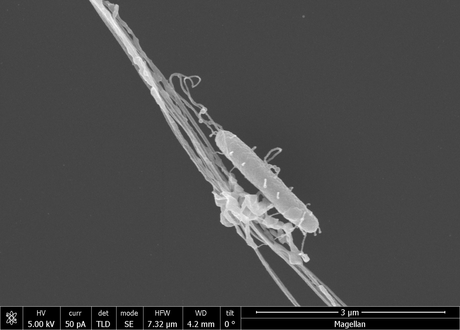
Pili-mediated attachment of a bacterium to a diatom spine revealed by scanning electron microscopy. Credits: Dr. Mathieu Forget